Home

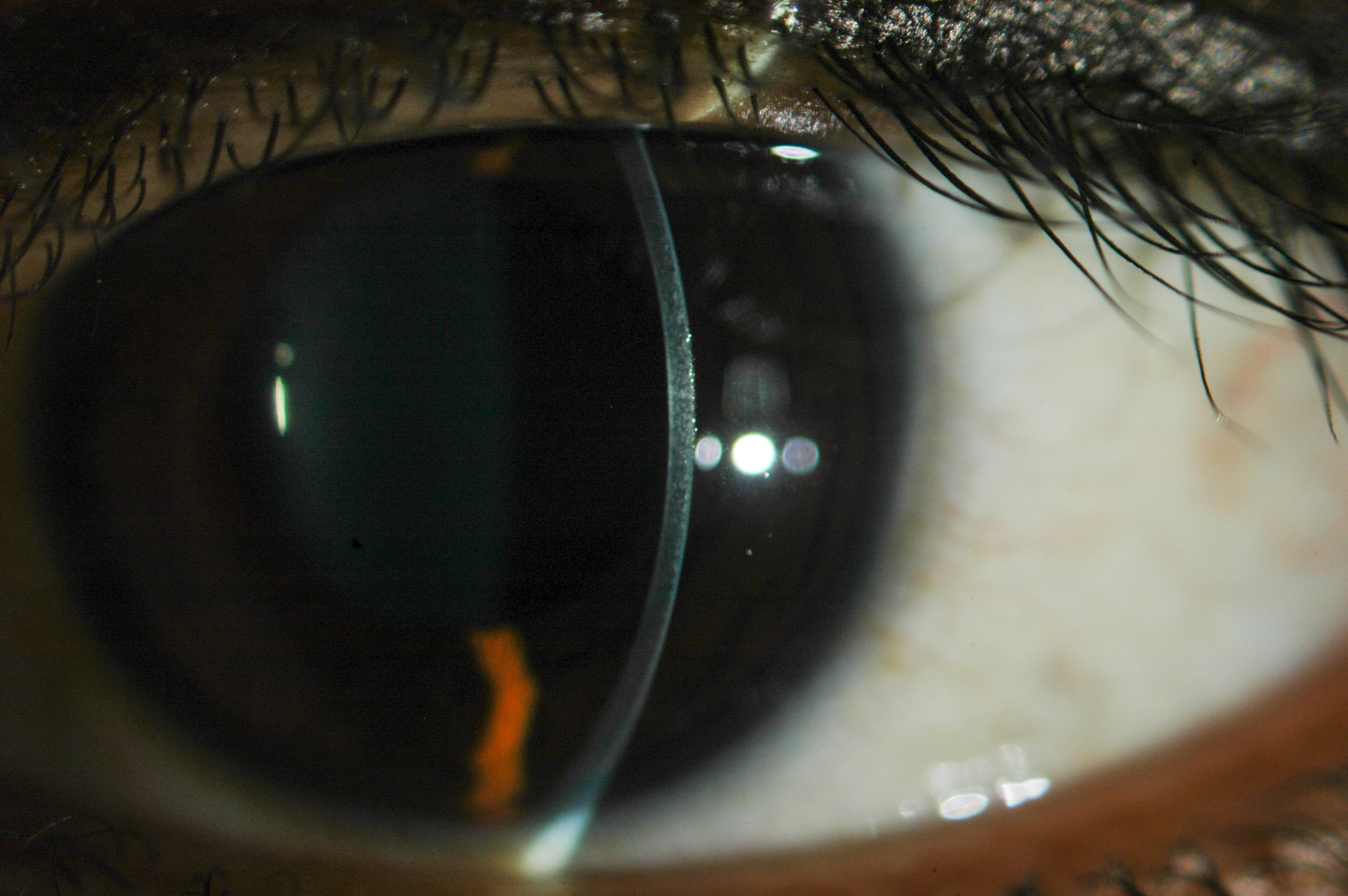
locken Reservoir Kunst cornea slit lamp Fortsetzen Kantine Stören

Slit-lamp Biomicroscopy

Slit-lamp exam Information | Mount Sinai - New York

Corneal Pachymetry and Endothelial Microscopy by Slit-Lamp | IntechOpen

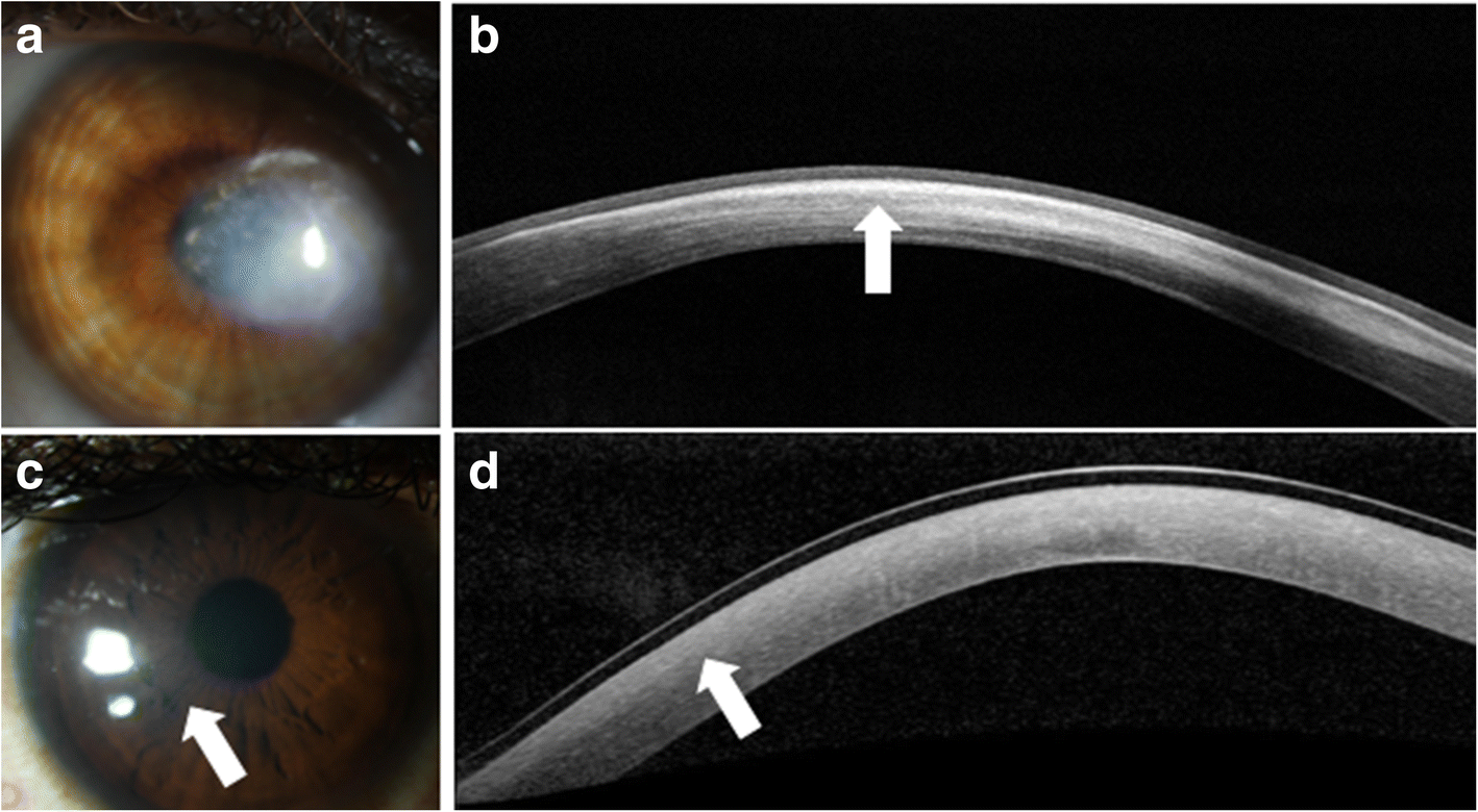
Optical coherence tomography for ocular surface and corneal diseases: a  review | Eye and Vision | Full Text
Optical coherence tomography for ocular surface and corneal diseases: a review | Eye and Vision | Full Text

Slit lamp images Flashcards | Quizlet

A, Slit-lamp biomicroscopy imaging of the cornea of this patient... |  Download Scientific Diagram
A, Slit-lamp biomicroscopy imaging of the cornea of this patient... | Download Scientific Diagram

How to Use a Slit Lamp - American Academy of Ophthalmology

Franceschetti Hereditary Recurrent Corneal Erosion - American Journal of  Ophthalmology
Franceschetti Hereditary Recurrent Corneal Erosion - American Journal of Ophthalmology

Short-term regression of corneal neovascularization with combination t |  IMCRJ
Short-term regression of corneal neovascularization with combination t | IMCRJ

Top tips for slit-lamp imaging - part 2 - Optician

Figure 1. [Findings on slit lamp examination of the cornea in cystinosis].  - GeneReviews® - NCBI Bookshelf
Figure 1. [Findings on slit lamp examination of the cornea in cystinosis]. - GeneReviews® - NCBI Bookshelf
Progenitor cells exist in the transitional zone between the peripheral  corneal endothelium and trabecular meshwork - EyeResearchNow.com
Progenitor cells exist in the transitional zone between the peripheral corneal endothelium and trabecular meshwork - EyeResearchNow.com

Corneal lens implant (Video) - TimRoot.com

A) Slit lamp photograph of the right eye showing superficial... | Download  Scientific Diagram
A) Slit lamp photograph of the right eye showing superficial... | Download Scientific Diagram

Cornea Abrasion Boise | Scratch on Cornea Idaho

Corneal ulcer, slit-lamp examination - Stock Image - C034/8023 - Science  Photo Library
Corneal ulcer, slit-lamp examination - Stock Image - C034/8023 - Science Photo Library

Cornea verticillata in Fabry disease: a comparative study between slit-lamp  examination and in vivo corneal confocal microscopy | British Journal of  Ophthalmology
Cornea verticillata in Fabry disease: a comparative study between slit-lamp examination and in vivo corneal confocal microscopy | British Journal of Ophthalmology

Slit-lamp photographs of the cornea in the left eye with chemical... |  Download Scientific Diagram
Slit-lamp photographs of the cornea in the left eye with chemical... | Download Scientific Diagram

Optic Section - A Study Guide to the Slit Lamp Biomicroscope

Ophthalmology Dx: Blurred Sight for Sore Eye - Ophthalmology Advisor

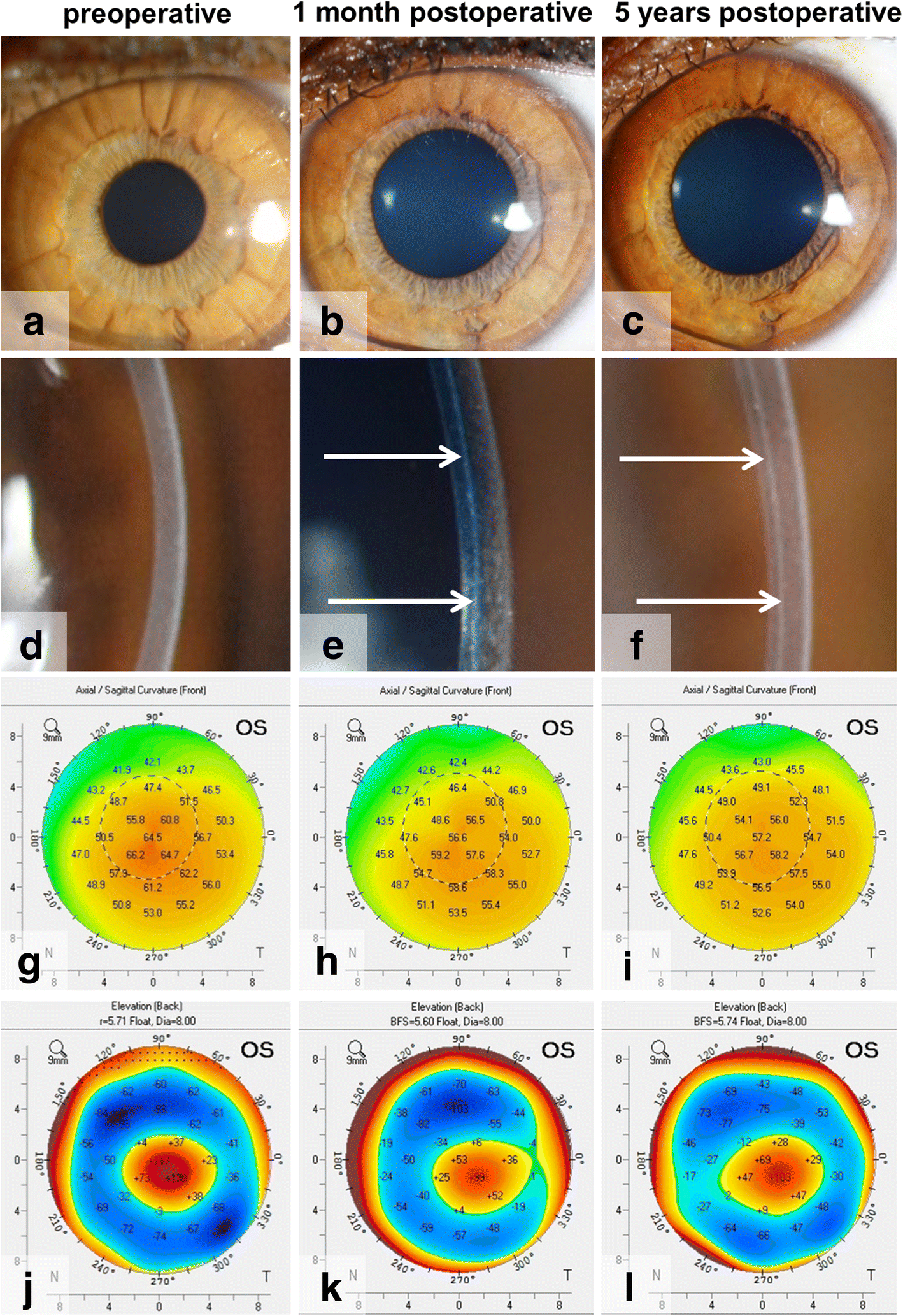
Bowman layer transplantation in the treatment of keratoconus | Eye and  Vision | Full Text
Bowman layer transplantation in the treatment of keratoconus | Eye and Vision | Full Text

Photographs of the cornea from six individuals examined using slit lamp...  | Download Scientific Diagram
Photographs of the cornea from six individuals examined using slit lamp... | Download Scientific Diagram

DLK scar under a lasik flap (Video) - TimRoot.com